The first 3D mapping of cortical sulcal patterns in autistic children
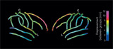
Courtesy of Jennifer G. Levitt, University of California, Los Angeles.
3D variability in cortical sulci between the patient and the control
groups.
Jennifer Levitt and
colleagues (Cortical sulcal maps in autism, Cerebral Cortex 13, 728-735,
2003)
using Magnetic Resonance Imaging (MRI) developed the first ever
three-dimensional mapping of cortical sulcal patterns in autistic disorder.
They studied autistic and normal children and built from single subject scans
high resolution average sulcal maps for the two groups. The results reveal
widespread deviations in cortical surface anatomy.
Among neuro-anatomical
abnormalities that accompany autism, there are differences in limbic,
cerebellar and neocortical regions, although there is no clear consensus on the
role played by those abnormalities, their connection with pathology is no more
in doubt. Jennifer Levitt’s group provides an important contribution to the
study of the neocortex affected areas. Examining the differences between
autistic and normal children in the pattern of cortical gyri and sulci could
provide a piece of evidence that leads to the solution of problems related to
the developmental pathology of the disorder. For example, the posterior
shifting of the inferior frontal gyrus in normal development, doesn’t take
place in its autistic counterpart, resulting in an anterior displacement. Such
differences, Levitt speculates, could reflect delayed or incomplete maturation
in the frontal lobe.
BM&L-October 2003